| Main centres: | 1-3 business days |
| Regional areas: | 3-4 business days |
| Remote areas: | 3-5 business days |
TECHNICAL SPECIFICATIONS:
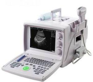
| Application range This device is suitable in hospitals and clinics for diagnosing liver, GB, spleen, kidney, pancreas, heart, bladder, and uterus.Feature set This device uses micro computer technology, Digital Scanning Converter (DSC), variable aperture, multi-section dynamic focusing, high dynamic low noise and wide band pre-amplifier, log compression, TGC control, dynamic filtering, edge enhancement and frame correlation. The embedded components allow a clear, stable and high resolution image. Front End with 80 arrays, 20 channels Display modes: B, B+B, B+M, M; Gray scales: 256 levels. Real time scanning or picture freezing for analysis. Image amplification, black-white reversed, up/down conversed and shift depth in Real Time. Soft-touching keyboard and track ball to make operation faster, convenient and flexible. Video connector, outputting PAL-D standard that can be easily connected to large external monitor, VCR or image printer. Portable structured with plastic cover. The use of switch power supply, integrate circuits (FPGA) and Surface Mounting Technology (SMT) makes this device portable with a small volume and light weight. This device has passed clinical verifications of safety and diagnosis validity.Technical Specification Standard probe : 3.5MHz Triple frequency Electronically convex Optional probe : 1, L40/7.5MHz Linear Probe 2, L40/7.5MHz EndoRectal Linear 3, R13/6.5MHz Transvaginal Convex Probe Scanning depth : 200mm Acoustic Power Mode M (C1-2/R60) :- P- : 1.0498mpa Iob 1.316mw/cm2 Ispta 2.63mw/cm2 Lateral resolution : 3mm (depth 80mm) or 4mm (80mm < depth 130mm) Axial resolution : 2mm (depth 80mm) or 3mm (80mm < depth 130mm) Dead zone : 5mm Geometric position precision (%) : Horizontal : 15 Environment requirements |